
Intraoral Cameras in Pine Beach, NJ
Our dentist may use an intraoral camera to look at your teeth. The wand-like instrument has a camera on the end that connects to a digital screen so that you can see what Dr. Mariya Medlenov sees during your treatment. To learn more about our intraoral cameras in Pine Beach, New Jersey, at Century Dental East, call 732-341-6010 to schedule your next appointment!
An intraoral camera is a tool we use to get a closer look at different parts of your smile. The intraoral camera is a small, pen-shaped device that we can use to take high-quality color photos of your teeth and gums, as well as other areas of your mouth as needed. Because the intraoral camera is so small and easily maneuverable, we can use it to see angles in your mouth that we cannot easily view with the unaided eye. We may utilize the intraoral camera to better evaluate your oral health and diagnose dental problems like tooth decay and periodontal disease, as well as tooth damage like a cracked or chipped tooth. The intraoral camera is also useful in following up after a treatment has been completed.
In addition to helping our team get a better view of your smile, the images taken with the intraoral camera allow you, our patient, to gain a better understanding of your oral health and dental treatments. We can also send these photos to other dentists and specialists and can submit them to dental insurance companies as needed. To learn more about the uses and benefits of intraoral cameras, we invite you to call or visit our dentist and team soon.
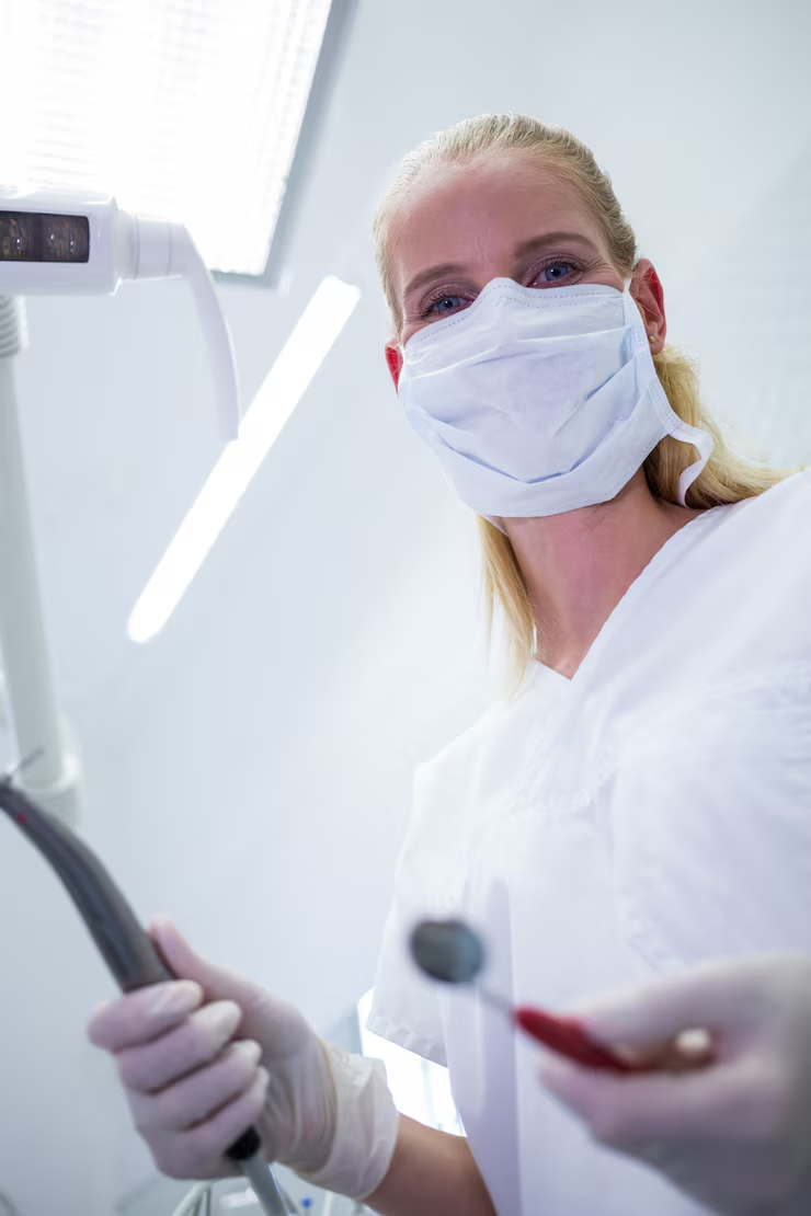
What an Intraoral Camera Shows You
The clearest explanation of a dental problem is usually a picture of your own tooth.
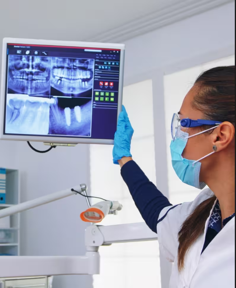
Seeing what the dentist sees
An intraoral camera is a small wand with a lens and light at the tip. It photographs teeth and gums from angles that are impossible to reach with a mirror and difficult to describe in words, then puts that image on a screen at chairside, enlarged many times over.
That changes the conversation. Instead of being told a back molar has a crack running through it, you look at the crack. Patients make treatment decisions more confidently when they can see the reason for the recommendation rather than take it on trust.

Cracked and chipped teeth. Hairline fractures show up clearly when magnified and lit.
Worn or leaking fillings. Gaps and staining at the margins of older restorations.
Gum condition. Inflammation, recession and areas of buildup along the gumline.
Soft tissue. Sores, discolored patches and irritation from an appliance or a sharp edge.
Hard-to-see angles. The back of the last molars and the tongue side of the lower teeth.
Catching Problems Early and Tracking Changes
A photograph taken today becomes the reference point for next year's visit.

Watching an area rather than guessing
Not every finding needs treatment straight away. A small area of wear, a patch of gum recession or a hairline crack may simply need watching. A dated image lets Dr. Mariya Medlenov compare that area at your next visit and see whether it has actually changed, instead of relying on memory or a written note.
Working alongside X-rays, not instead of them
The camera shows surfaces. It cannot see through enamel or below the gumline, which is why it works together with digital dental X-rays rather than replacing them. Between the two, most of what matters becomes visible: the camera for the outside, radiographs for what sits underneath. Both are part of the dental technology we use in Pine Beach.
Before and after. Images taken at the start and end of treatment show exactly what was done.
Home care that lands. Seeing plaque along a specific gumline is more useful than a general reminder to brush better, and gives your dental cleaning visits a clear focus.
Shared records. Images can be sent to a specialist or submitted with an insurance claim to document what was found.
What the Appointment Is Like
A minute or two, with nothing to feel.
How the images are taken
The camera tip is about the size of a pen and is covered with a fresh disposable sleeve for each patient. It is moved around the mouth, resting lightly against the teeth, while images are captured of the areas we want to look at closely. There is no discomfort, no numbing and no radiation involved. Most patients find it easier than having a routine X-ray taken.
During an exam.
Images are usually taken as part of a dental exam, when something specific needs a closer look.

When something hurts.
If a tooth is sensitive or a filling feels rough, the camera often finds the cause quickly. For a broken tooth or lost restoration, see emergency dental care.
Reviewing together.
The images stay on the monitor while we talk through the options, so you can ask questions with the picture in front of you.
If you gag easily.
Say so. Positioning can be adapted, and nitrous oxide helps some patients relax through it.
Why It Matters for Your Care

Better information, clearer decisions
Dentistry involves a lot of choices that are easier to make when the problem is visible. Whether to repair a worn filling now or watch it, whether a crack has reached the point where a crown is the safer option, whether an area of gum irritation is settling. Photographs turn those from abstract judgments into something you can weigh up yourself.
Fewer surprises. You see a problem developing instead of hearing about it once it needs urgent work.
A shared record. Your images stay in your chart and travel with your file if you ever move practices.
Clearer claims. Documentation supports what is submitted to your plan; our financial options page explains how we handle the paperwork.
See your smile for yourself in Pine Beach
Century Dental East serves patients throughout Ocean County from our office on Washington Avenue in Pine Beach. To book an exam with Dr. Mariya Medlenov, call (732) 341-6010 or contact us online.
Visit Our Pine Beach Office
Century Dental East: Gentle, Modern Dentistry for Families in Pine Beach, NJ.
241 Washington Avenue, Pine Beach, NJ 08741
(732) 341-6010


Hours
Mon 9:00 a.m. to 7:00 p.m.
Tue 9:00 a.m. to 7:00 p.m.
Wed 9:00 a.m. to 5:00 p.m.
Thu 9:00 a.m. to 5:00 p.m.
Fri-Sun Closed
Serving patients across
- Toms River
- Lakewood
- Lakehurst
- Forked River
- Bayville
- Beachwood
- Pine Beach
- Barnegat Light
- Ocean Gate
- Maple Shade
- Leisure Village
- Cedar Grove
- Waretown
- Howardsville
- Brookville
- Holly Park
- South Toms River