
Digital Dental X-Rays in Pine Beach, NJ
Dr. Mariya Medlenov uses digital X-rays to see the state of your teeth below the gumline in order to see what is the best treatment to give you based on your needs. Once Dr. Medlenov does so, our dentist can help get your oral health back on track! To schedule your next appointment at Century Dental East or to learn more about the digital X-rays we use in Pine Beach, New Jersey, call 732-341-6010!
Digital X-rays are one of the advanced diagnostic tools we use to provide you with the best possible care. Digital radiography has changed the way we take dental X-rays by making the process faster, more comfortable and more convenient than ever before.
Digital X-rays use a digital sensor to take images of your teeth, unlike traditional X-rays that use film. Digital X-rays use significantly less radiation than conventional film X-rays and require the use of no chemicals for developing the images.
Our dentist can then instantly view the high-quality images on monitors right in the treatment room to provide you with efficient, accurate diagnoses and care. Our team is able to use digital radiography to identify and diagnose several types of dental problems, such as:
- Decay in between teeth
- Developmental abnormalities
- Improper tooth root positioning
- Cysts and abscesses
- Fractures in existing fillings
- Tumors
- Infection in the tooth nerves
- Bone loss
Digital dental X-rays are used to plan many kinds of dental treatment, such as oral surgery procedures, root canal treatment, dental implants, orthodontics and more. To learn more about digital radiography and how we use X-rays at our office, we welcome you to call or visit us soon.
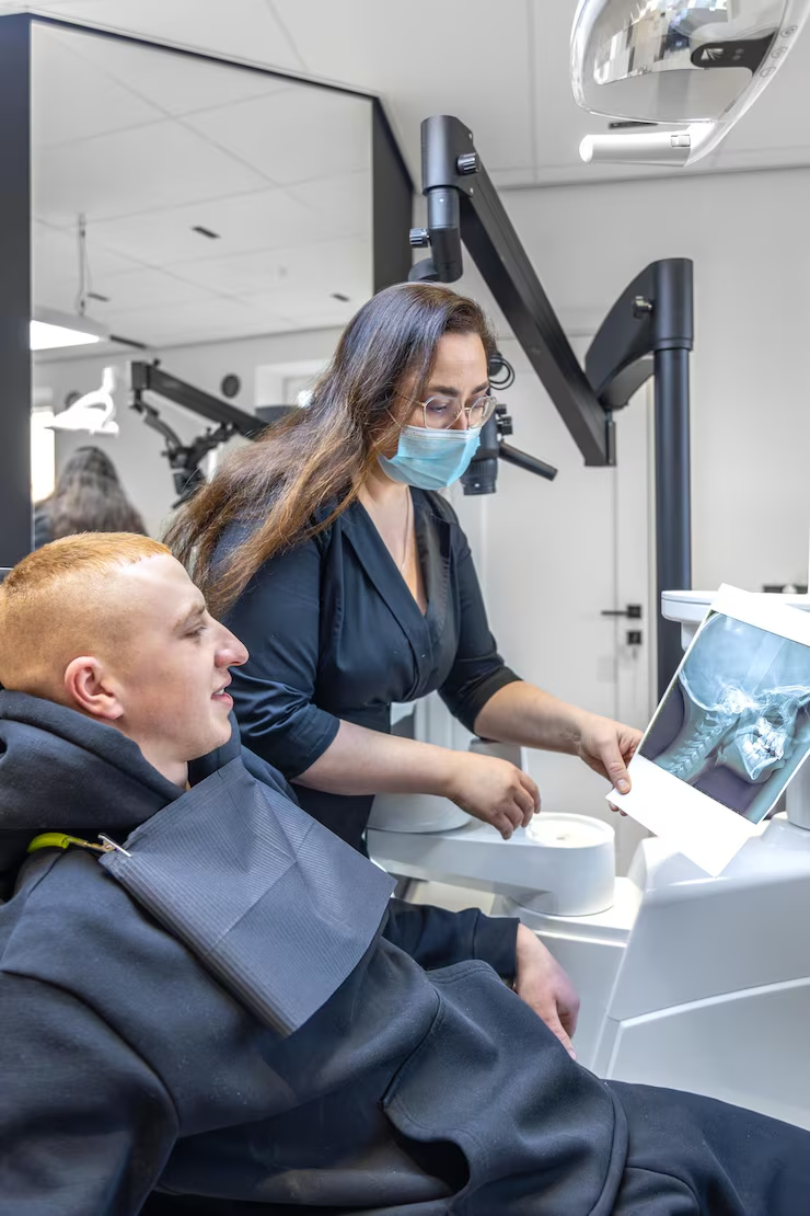
What Dental X-Rays Show That an Exam Cannot
A visual exam covers the surfaces we can see. Roughly a third of each tooth, and everything supporting it, sits out of sight.
Below the gumline and between the teeth
Two of the places decay starts most often are the tightly contacting surfaces between neighboring teeth and the area beneath an existing filling. Neither is visible to the eye or reachable with a probe. Radiographs make those areas readable, along with the bone that holds each tooth in place and the root structures below.
Bite-wing images.
Show the crowns of upper and lower back teeth together, which is how decay between teeth is usually caught.

Periapical images.
Show a whole tooth from crown to root tip, used when a specific tooth is painful or has been injured.
Panoramic images.
Show the full upper and lower jaw in one view, useful for wisdom teeth, jaw joints and eruption patterns in children.
For treatment planning that needs three-dimensional detail, such as implant placement, our CBCT scanner provides a different kind of image again. All of it sits within our wider dental technology at the Pine Beach office.
Catching Problems Early
Nearly every dental problem is smaller, simpler and less uncomfortable to treat when it is found early.

Small now, or larger later
A spot of decay between two molars is a small filling if it is found at a routine visit. Left until it causes pain, the same cavity may have reached the nerve, and the treatment becomes root canal therapy and a dental crown. Radiographs are how the first version of that story gets caught.

Decay between and beneath. Cavities forming in contact areas and under existing restorations.
Bone loss. Changes in the bone level around teeth, which is how gum disease is tracked over time.
Infection. Abscesses and cysts at the root tip, often before there is any pain.
Failing dental work. Fractures or gaps at the edge of an older filling.
Development and position. How permanent teeth are forming in children, and whether wisdom teeth are angled to cause trouble.
This is why images are taken at intervals alongside your dental exam, rather than only when something hurts.
Digital X-Rays Compared With Film
The same diagnostic purpose, handled with a sensor instead of a sheet of film.
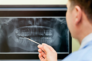
What changes for you in the chair
With digital radiography, a small electronic sensor replaces the film packet and the image appears on the monitor within seconds. There is no darkroom step and no waiting between images, so the whole sequence takes less time. Digital X-rays also use less radiation than traditional film X-rays, and they use no developing chemicals at all.
Immediate images. Dr. Mariya Medlenov can review them with you in the room, on screen, at the same visit.
Adjustable on screen. Images can be enlarged and the contrast altered, which helps in reading fine detail.
Easy to compare. Stored images sit side by side with older ones, so slow changes in bone level or a suspect area can be tracked year to year.
Simple to share. Images can be sent to a specialist or an insurer without copying film.
Radiation exposure is kept as low as reasonably achievable: a lead apron and thyroid collar where appropriate, images taken only at intervals your dental history justifies, and each one taken for a specific reason. Tell us if you are pregnant or may be, and we will discuss what can safely be deferred.
What the Appointment Is Like
A few minutes, and nothing to feel
Taking dental X-rays is quick and painless. You sit in the chair, a protective apron is placed, and the sensor is positioned in your mouth for each image. You are asked to hold still and bite gently for a moment, and the exposure itself lasts a fraction of a second. A full set of bite-wings takes only a few minutes.
If you gag easily. Tell us. Positioning can be adjusted, and nitrous oxide helps some patients get through imaging comfortably.
For children. Smaller sensors are used, and the number of images is kept to what is actually needed.
Transferring records. If you have had recent X-rays elsewhere, we can often request them rather than repeat them. Ask when you book.
Coverage. Diagnostic images are covered by most dental plans at set intervals; see our financial options page for how we handle billing.
Schedule an exam in Pine Beach
Digital dental X-rays are part of thorough care at Century Dental East, serving patients across Ocean County. Call (732) 341-6010 or contact our office to schedule with Dr. Mariya Medlenov.
Visit Our Pine Beach Office
Century Dental East — Gentle, Modern Dentistry for Families in Pine Beach, NJ.
241 Washington Avenue, Pine Beach, NJ 08741
(732) 341-6010


Hours
Mon 9:00 a.m. – 6:00 p.m.
Tue 9:00 a.m. – 7:00 p.m.
Wed 9:00 a.m. – 5:00 p.m.
Thu 9:00 a.m. – 5:00 p.m.
Fri-Sun Closed
Serving patients across
- Toms River
- Lakewood
- Lakehurst
- Forked River
- Bayville
- Beachwood
- Pine Beach
- Barnegat Light
- Ocean Gate
- Maple Shade
- Leisure Village
- Cedar Grove
- Waretown
- Howardsville
- Brookville
- Holly Park
- South Toms River